OncoVision — Cancer Residual Detection System
Intraoperative fluorescence with AI overlays to expose residual malignant tissue in real time. Multi-spectral capture, tumour-targeted agents and CV segmentation guide complete excision and reduce re-ops.
Overview
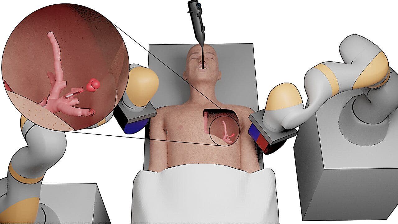
OncoVision is an advanced intraoperative imaging and analysis platform designed to help surgeons identify residual malignant tissue in real time during oncologic procedures. The system leverages tumour-targeted fluorescent agents that selectively bind to cancerous cells, which then emit a detectable signal under specific excitation lighting. This optical signal is captured using a fluorescence-sensitive camera and processed through a multi-spectral pipeline to enhance signal-to-noise and quantify malignancy likelihood. On top of this, computer-vision and deep-learning models generate overlays that outline or colorize regions suspected to harbor residual disease. These overlays are rendered directly on the live surgical feed so that the operative field and AI guidance are available in a single, familiar visual context. By providing immediate, data-backed cues to help delineate margins and locate otherwise subtle remnants, OncoVision helps ensure complete tumor excision, thereby reducing recurrence rates and improving patient outcomes.
Clinical Problem
Intraoperative decision-making in oncologic surgery often relies on the surgeon’s visual judgement and tactile feedback, supplemented by frozen section histopathology when available. However, microscopic residual disease can be missed, particularly when tumor margins are irregular or located in anatomically complex regions. Waiting for pathology adds time and may still fail to capture every positive margin. When residual cancer remains, patients face higher chances of local recurrence, re-operations, additional adjuvant therapy, and increased morbidity. The clinical challenge is to provide the surgeon with a sensitive, immediate indicator of tissue suspicious for malignancy without disrupting established workflow or adding unsafe latency. A system that can illuminate residual disease at the point of care — accurately and in real time — can significantly improve completeness of resection.
Methodology
- Administer tumour-targeted fluorescent agent preoperatively or intraoperatively, ensuring sufficient uptake by malignant tissue.
- Illuminate the operative field with appropriate narrow-band excitation and capture the resulting emission using a high-sensitivity camera.
- Process the multi-spectral stream using AI/CV to enhance contrast, suppress noise, and assign malignant probability maps.
- Render color-coded overlays on the live feed with adjustable opacity so surgeons can view anatomy and AI guidance together.
- Integrate with existing theatre cameras/monitors to preserve ergonomics and avoid latency beyond clinical tolerance.
Tech Stack
Equipment
Expected Outcomes
Optics bench + segmentation prototype in progress; simulation trials precede clinical validation with oncology partners.