Skip the 3D story
ManMed robotic stethoscope concept image

01 · Robotic Auscultation

A complete robotic auscultation bench

A myCobot 280 carries a stethoscope chestpiece to a synthetic thoracic phantom. A short tube feeds ManMed’s acoustic sensing module, which streams the signal over a wired link to an AI workstation and display.

Research set-up visualisation · synthetic phantom, no patient data.

02 · Platform

myCobot 280 — six joints, 280 mm reach

Elephant Robotics’ desktop six-axis arm: 280 mm working radius, 250 g rated payload and ±0.5 mm stated repeatability — light enough for a bench, with a payload budget that suits a chestpiece but not an electronics box.

Third-party hardware, modelled approximately from published dimensions and joint ranges (myCobot 280 for M5). The exact variant used by ManMed is to be confirmed.

03 · End effector

A lightweight chestpiece mount

An adapter plate on the J6 flange, a spring-loaded compliant mount and a clamp ring hold a 44 mm diaphragm chestpiece. Only the chestpiece and a short tube ride on the wrist — the electronics stay on the bench.

The compliant mount is a conceptual design.

04 · Acquisition

Approach, contact, hold, listen

The arm visits auscultation areas on the phantom, lowers the diaphragm until it is in contact, and holds still while the module records.

Motion is an inverse-kinematics visualisation, not a validated robot controller. Sites and force values are illustrative.

05 · Acoustic path

A short tube to the sensing module

Sound travels through the tube into a coupler chamber with an electret microphone. The enclosure carries the tube inlet, a USB-C data port, DC input, ventilation slots and mounting flanges.

Enclosure layout is illustrative.

06 · Electronics

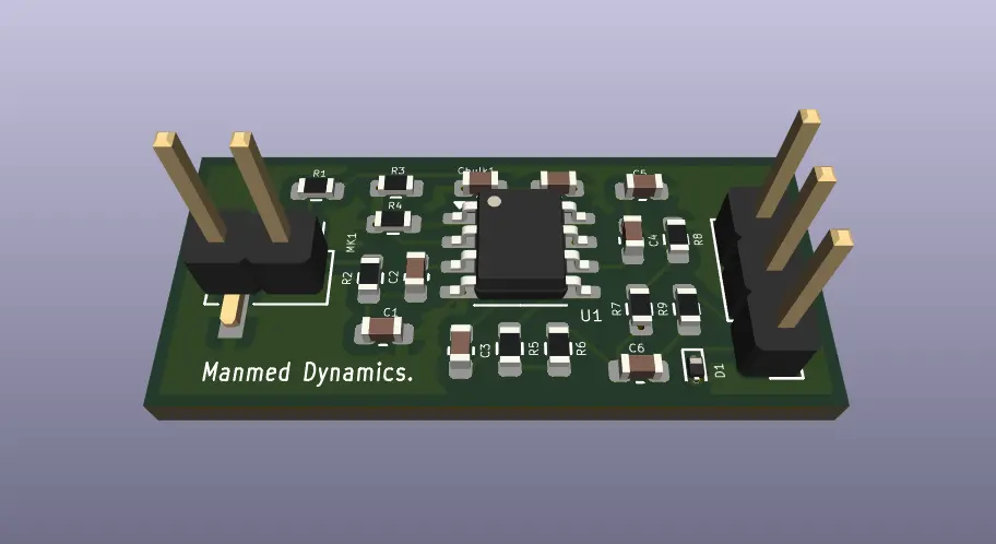
ManMed’s 10 × 24 mm preamplifier

A two-layer board built around the OPA1678 low-noise op-amp: an electret-microphone interface, a preamplifier and a filter chain designed for the ≈20 Hz – 1.2 kHz band where heart sounds lie. Wired by design — no Bluetooth module.

PCB design stage (schematic, layout, DRC and Gerbers complete). Fabrication, simulation and measured performance are pending — no measured results are shown. The acquisition/USB board is conceptual.

07 · Processing

Wired to an AI workstation

A USB link carries the digitised audio to a liquid-cooled AI workstation for filtering, segmentation and analysis; the same workstation commands the robot over USB.

The workstation shown is an illustrative, unbranded design, not a specific product.

08 · Display

Synchronised signal views

Raw microphone signal, a band-passed phonocardiogram, a separate ECG channel, the spectrum and recording status on one research display.

All signals are synthetic. The ECG channel is illustrative — no ECG acquisition hardware is part of this set-up.

09 · Engineering view

One system, end to end

Robot → chestpiece → tube → acoustic module → workstation → display, as a technical drawing.

10 · Robotic Auscultation

Repeatable placement, a dedicated signal chain

ManMed’s robotic auscultation research: consistent chestpiece contact on a robot platform and a low-noise, wired acquisition path.

Research detail →

Research programme

Robotic Stethoscope — Force-Controlled Robotic Auscultation Module

In progress

Research & IP concept — aligned with ManMed IP

  • Robotics
  • Acoustics
  • Signal processing

Overview

A precision auscultation module designed for robotic platforms — consistent contact force, noise rejection, and high-fidelity signals for clinical decision support.

The module is conceived as a robot end-effector with constant contact force, a denoising pipeline and clinical signal metrics, aligned with ManMed IP. ManMed has designed a dedicated acoustic sensing / pre-amplifier board for the module.

Clinical problem

Manual auscultation quality depends on how the chest piece is placed and how firmly it is held: inconsistent contact pressure and handling noise change what is heard. A robotic platform needs an auscultation end-effector that applies repeatable contact force and delivers clean, comparable acoustic signals.

Methodology

  1. 01

    Mount the chest piece on a compliant, force-controlled end-effector so contact pressure stays consistent.

  2. 02

    Measure contact force continuously and regulate it at the robot interface.

  3. 03

    Acquire the acoustic signal through a dedicated microphone pre-amplifier board.

  4. 04

    Apply filtering and denoising, then compute signal metrics for clinical decision support.

Technical explanations

Why a robot holds the chestpiece

What a clinician hears depends on where the chestpiece sits and how firmly it is pressed. A robot can return to the same location with the same contact conditions, which is the starting point for comparable recordings over time.

The myCobot 280 platform

A six-axis desktop arm with a 280 mm working radius and a 250 g rated payload (Elephant Robotics’ figures for the myCobot 280 for M5). The small payload is why only a chestpiece and a short tube are mounted on the wrist, while the electronics stay on the bench.

Acoustic path

Heart sounds are carried from the diaphragm through a short tube to a coupler chamber in front of an electret microphone, keeping the sensitive electronics away from the moving arm and from handling noise.

The pre-amplifier board

ManMed’s 10 × 24 mm two-layer board pairs an electret-microphone interface with an OPA1678 low-noise op-amp and a filter chain designed for the roughly 20 Hz – 1.2 kHz band where heart sounds lie, with a solid ground plane and noise-aware routing.

Wired, not Bluetooth

The design deliberately leaves out wireless transmission, unlike many commercial digital stethoscopes, to keep the clinical data path simple and contained.

Processing and display

The digitised signal is filtered, denoised and segmented into heart-sound events (S1/S2) on a workstation; the display on this page uses synthetic signals to explain those steps.

Limitations

  • No patient recordings, clinical results or diagnostic claims are shown; every waveform on this page is synthetic.
  • The pre-amplifier PCB is at the design stage: fabrication, simulation and measured noise or frequency response are pending.
  • The ECG channel on the display is illustrative — no ECG acquisition hardware is part of the depicted set-up.
  • The myCobot model is an approximation from published dimensions; the module enclosure, acquisition board and workstation are illustrative.

Stack & equipment

Technology

  • Force control
  • Acoustic sensing
  • Denoising pipeline
  • Signal metrics
  • KiCad (PCB design)

Equipment

  • Elephant Robotics myCobot 280 — six-axis desktop arm (third-party platform; exact variant to be confirmed)
  • Stethoscope chestpiece on a lightweight end-effector mount (compliant mount conceptual)
  • Microphone pre-amplifier PCB (ManMed design): 10 × 24 mm, 2-layer, OPA1678 op-amp, electret microphone interface
  • Wired acquisition to a processing workstation and display (illustrative)

Status & next steps

Patent concept and engineering research. The pre-amplifier PCB design is complete (schematic, layout, design-rule check, fabrication files); fabrication, simulation and measured performance are pending. Clinical performance has not been published.

Content migrated from https://manmed.in/ (Featured Research) and https://manmed.in/projects (Thesis track). 3D models are illustrative engineering visualisations.

Robotic Auscultation — image from the ManMed research archive
Robotic Auscultation · ManMed research archive